Our latest product is for preclinical MRI
Introducing Metis
by MR Shim GmbH
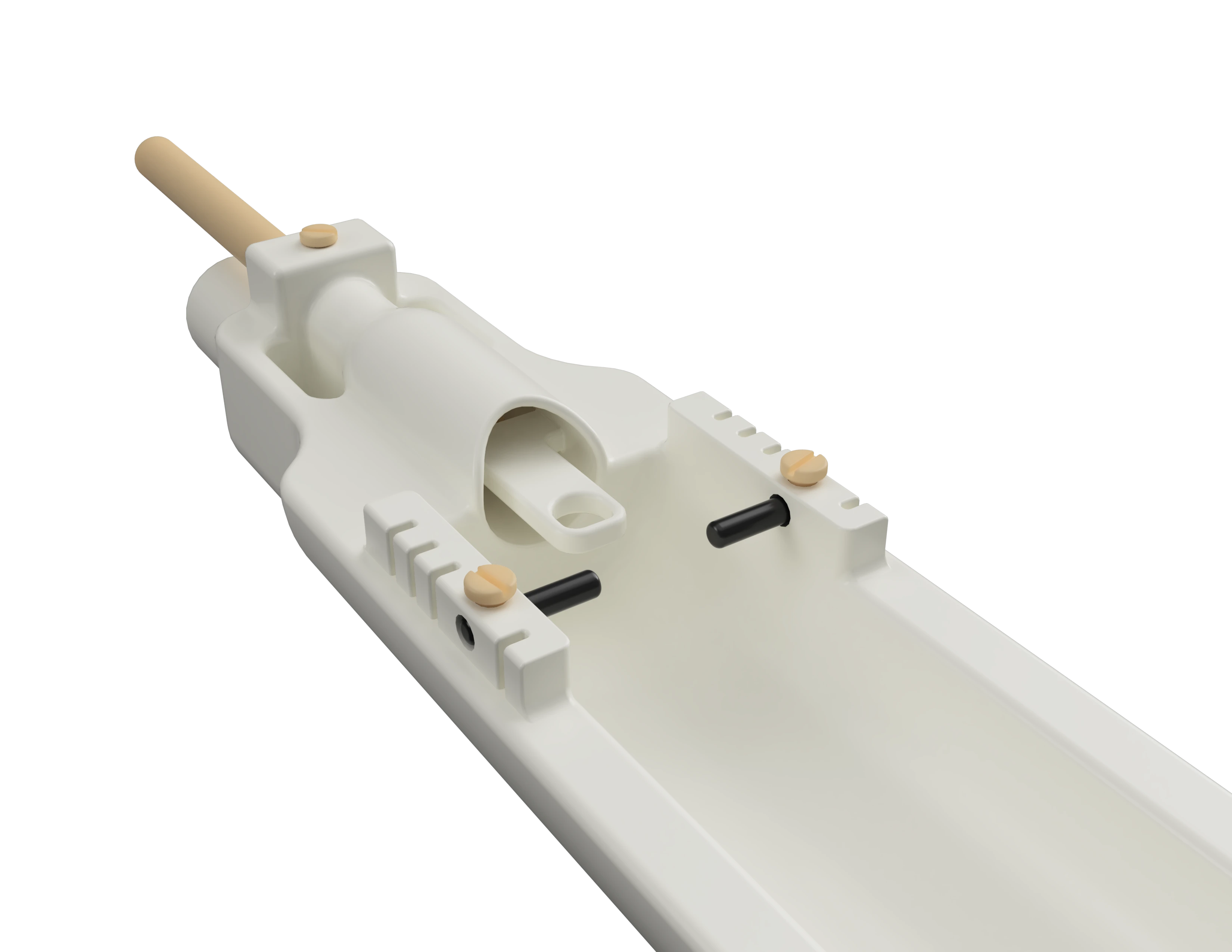
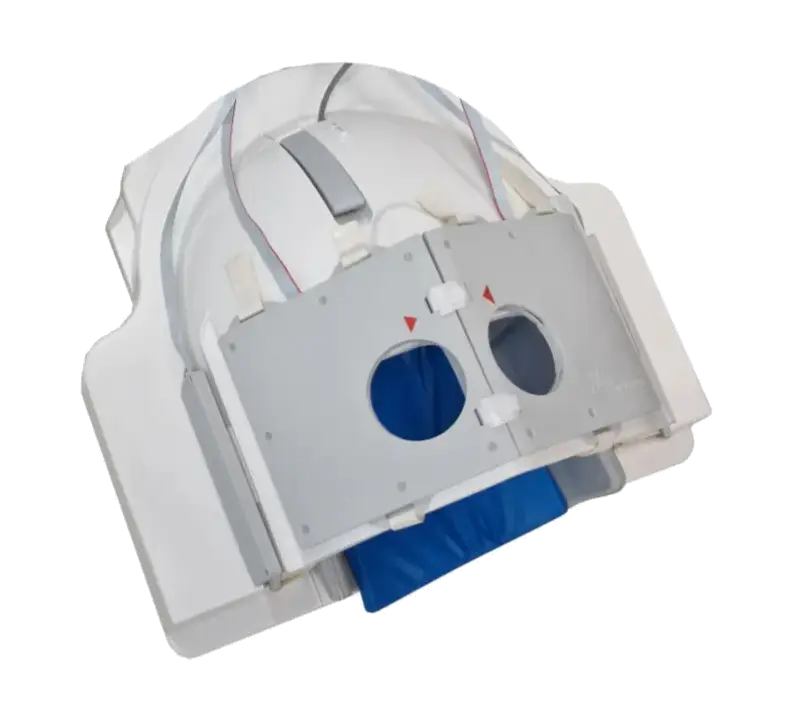
An innovative small animal cradle with integrated local shim coils engineered for optimal B0 shimming in preclinical MRI: Recovering signal dropouts, and eliminating susceptibility artifacts and distortions. (Also available as an add-on for existing cradles).

Our research is supported by:


Loading...
Recover signal in pre-frontal, temporal and deep brain areas.
2D EPI
Siemens Prisma 3T
TE=30ms; 25 x 25 x 25 mm

Reduce MRI scan repetitions and enhance data retention and utilisation with robust scan acquisitions.

Make groundbreaking scientific DISCOVERIES with higher signal-to-noise ratio (SNR) and complete signal recovery.

Leverage DYNAMIC and real-time shimming to correct for physiological changes and advanced field homogenisation techniques.


WIDE-VARIETY of products for neuro- and body-imaging applications in humans and small animals at all field strengths (0.05T to 11.7T).
How it works

Use Cases
Echo Planar Imaging
Echo planar imaging (EPI) enables us to capture the dynamic activities of the brain and unveil intricate patterns of neural activation. Poor field inhomogeneity can cast many image artifacts and sometimes even cause complete lack of signal in the EPI images. Improving the shim quality directly during acquisition can increase BOLD sensitivity, bring signal void back and reduce distortions....

Preclinical Imaging
Preclinical magnetic resonance imaging (MRI) is essential for enhancing our understanding of biological processes and disease mechanisms. It provides in-depth anatomical and functional information about biological samples, such as small animals, prior to human testing. This technique enables researchers to explore the underlying mechanisms of diseases, assess treatment effectiveness, and examine...

Imaging Near Implants
In recent years, there has been a significant increase in the number of individuals with implants in their bodies. MRI is often essential for post-operative assessment, monitoring of co-morbidities, and evaluation of surrounding tissues. While many modern implants are labeled as MRI-compatible, there remains an inherent issue of magnetic field inhomogeneity...

Quantitative Susceptibility Mapping
QSM is a technique used to non-invasively measure tissue susceptibilities, a key indicator in many disorders. Poor MRI magnetic field homogeneity, not unlike a distorted lens, can cast artifacts across the images such as streaking and blurring, incorrect quantification, distorted local field maps, and erroneous phase wraps...

Spectroscopic Imaging
The accurate assessment of brain metabolites using Magnetic Resonance Spectroscopy and Spectroscopic Imaging (MRS/MRSI) plays a crucial role in various clinical and research applications. Accurate B0 shimming especially at higher MRI field strengths is paramount for obtaining reliable metabolic information...

Lipid Suppression by Shaping Magnetic Fields
The presence of lipid signals surrounding the tissue of interest can significantly interfere with MRI image quality and contrast. Lipids produce a bright signal that hampers the visualization of other structures and may contaminate the signals from metabolites. The lipid signals can be suppressed directly during acquisition with an elegant hardware solution to reduce the burden on the imaging sequence and enhance data quality...

Prostate Imaging
Since a prostate MRI may save the patients from an unnecessary biopsy, any attempt to improve the MRI data quality could be highly impactful. B0 field homogeneity plays a vital role in generating high-quality MRI images of the prostate gland. Advanced shimming methods can help mitigate susceptibility-induced artifacts, such as distortion and blurring, resulting in improved image clarity and data accuracy...

Combined RF/Shim Coil
Ultra-high field (UHF) MRI is pushing the borders of medical imaging, offering unprecedented spatial resolution and exquisite image details. Advanced B0 shimming and RF technologies are arguably the two pivotal technologies essential for unlocking the full potential of UHF MRI. The combination of these technologies in one imaging coil enhances the usability and unlocks the full potential of UHF MRI...

Our Products
Didn't find what you were looking for?
We provide a wide range of shimming services and customisations to existing B0 shim systems upon customer request.
Custom specifications
Integrated RF/shim coil, stronger amplifiers, etc.
Passive shimming
Simulation and consultation for passive shimming
Body region
Tailored shim systems for specific applications
Field strengths
Customisable to any field strength and MRI scanner
Small animals
Local shim array for small animal imaging